

Skin Substitutes
A Swiss surgeon, Jaques Louis Reverdin, has been credited as performing the first skin autograft, in 1869, using a ‘pinch-graft’ technique. Although procurement methods have evolved, the split-thickness autograft remains the standard of care. Although reliable in experienced hands, this definitive solution to the open wound has substantial drawbacks, particularly donor site morbidity and availability. Unfortunately, despite decades of work, currently available alternatives are even more imperfect. A reliable permanent skin substitute will revolutionize the care of patients with burns and other difficult wounds.
The Perfect Skin Substitute Criteria for Autologous Split-Thickness Skin Would be:
Prevents water loss
Barrier to bacteria
Inexpensive
Long shelf life
Can be applied in one operation
Does not become hypertrophic
Flexible
Conforms to irregular wound surfaces
Can be used ‘off the shelf’
Does not require refrigeration
Cannot transmit viral diseases
Does not incite inflammatory response
Durable
Easy to secure
Grows with a child
Advances in Wound Closure
Cultured Epidermal Cells (CEA)
Dermal Analogs
Composite Substitutes
Cultured epidermal autograft (CEA)
Cultured epidermal autograft (CEA) is an important tool in the management of patients with massive burns. In full-thickness burns involving more than 90% TBSA it may be the only choice, given that procurement of the uninvolved skin will not be sufficient to cover the patient’s wound.
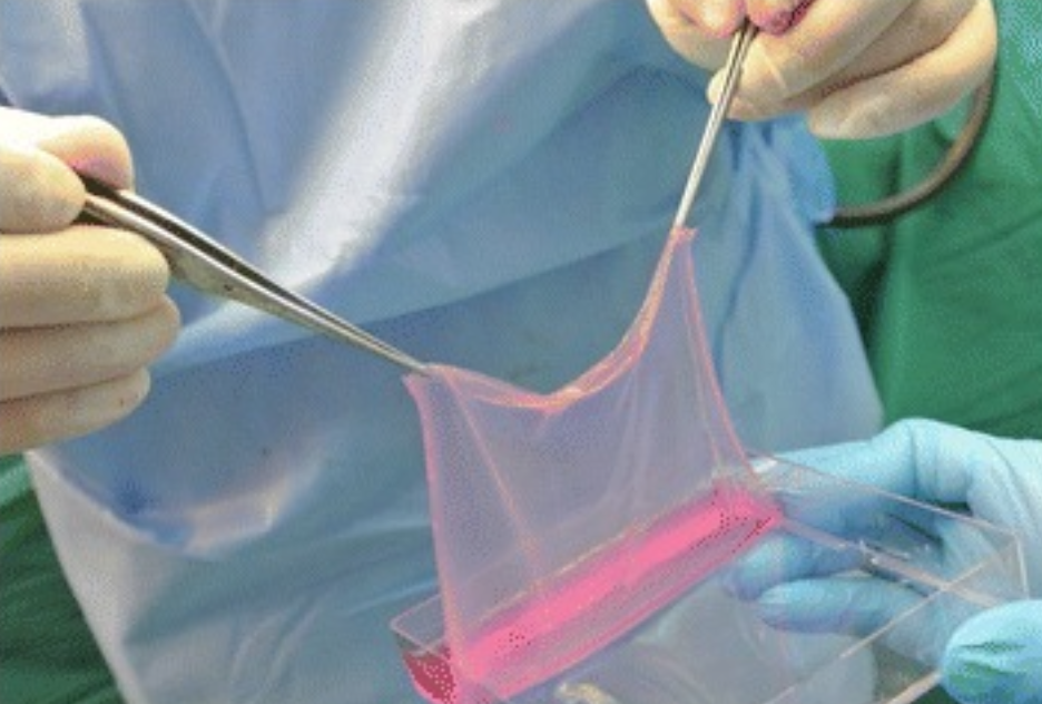
For over 20 years it has been possible to culture vast numbers of epithelial cells from a small skin biopsy, and this has led to the widespread clinical use of cultured epithelial grafts to cover burn wounds. Epithelial cells are procured from a full thickness skin biopsy, the cells being separated with trypsin. The resulting epithelial cell suspension is cultured in medium containing fetal calf serum, insulin, transferrin, hydrocortisone, epidermal growth factor, and cholera toxin, overlying a layer of murine fibroblasts that have been treated with a nonlethal dose of radiation that prevents them from multiplying. Colonies of epithelial cells expand into broad sheets of undifferentiated epithelial cells. These cells are separated from the culture vessel with trypsin and taken to secondary culture using the same techniques until confluent thin sheets of undifferentiated cells are obtained. The resulting sheets are removed from the dishes after treatment with dipase, which digests the proteins attaching the epithelial cells to the dish. The sheets of epithelial cells are attached to a petrolatum gauze carrier to ease handling.
The final product takes approximately 3 weeks to be ready for grafting and consists of sheets of keratinocytes, 2–8 cells thick.
When epithelial cultures were first used in patients with large burns it was hoped that they would provide the definitive answer to the clinical problem of the massive wound. With more frequent use of epithelial grafts, specific liabilities have become apparent including suboptimal engraftment rates and long-term durability. However, when faced with a very large wound and minimal donor sites, epithelial cell wound closure is a useful adjunct to split-thickness autograft, their liabilities and expense becoming more acceptable as wound size increases.
Many of the imperfections associated with epithelial cell wound closure may be attributed to the absence of a dermal element. Epithelial grafts are now commercially available. Application is generally most successful in wounds from which vascularized allograft has been removed. Despite scattered cases, application of cultured epithelial grafts onto synthetic dermal analogs has not been shown effective.
Source: Herndon, D. N. (2018). Total burn care. Edinburgh: Elsevier.